Introduction: A Game-Changer in Early Detection
introduction:-a-game-changer-in-early-detectionImagine a patient named Mr. Lee, a 50-year-old man with no obvious symptoms, who underwent a routine abdominal ultrasound at his doctor's recommendation. The scan revealed early signs of liver disease, a condition that could have remained undetected until it became much harder to treat. Thanks to this early detection, Mr. Lee was able to start treatment immediately, preventing severe complications and improving his long-term prognosis.
Ultrasound scans are one of the most powerful diagnostic tools in modern medicine, helping healthcare providers catch health problems early, often before symptoms even appear. By offering a non-invasive and radiation-free method to look inside the body, ultrasound plays a critical role in diagnosing a range of conditions—from heart disease and cancers to fetal abnormalities and organ dysfunction. Early diagnosis is one of the most effective ways to improve treatment outcomes, and ultrasound is helping to make this possible on a large scale.
In this article, we’ll explore the multifaceted role of ultrasound technology in modern healthcare. From its life-saving applications in early cancer detection to its importance in prenatal care, ultrasound continues to play a crucial role in improving patient health and saving lives.
Understanding Ultrasound Technology: More Than Just Sound Waves
understanding-ultrasound-technology:-more-than-just-sound-wavesAt first glance, ultrasound technology may seem simple, but it has evolved significantly over the years. Originally used for basic imaging, ultrasound now boasts several advanced features, such as 3D imaging, contrast-enhanced ultrasound, and AI-powered analysis, which further enhance its diagnostic capabilities.
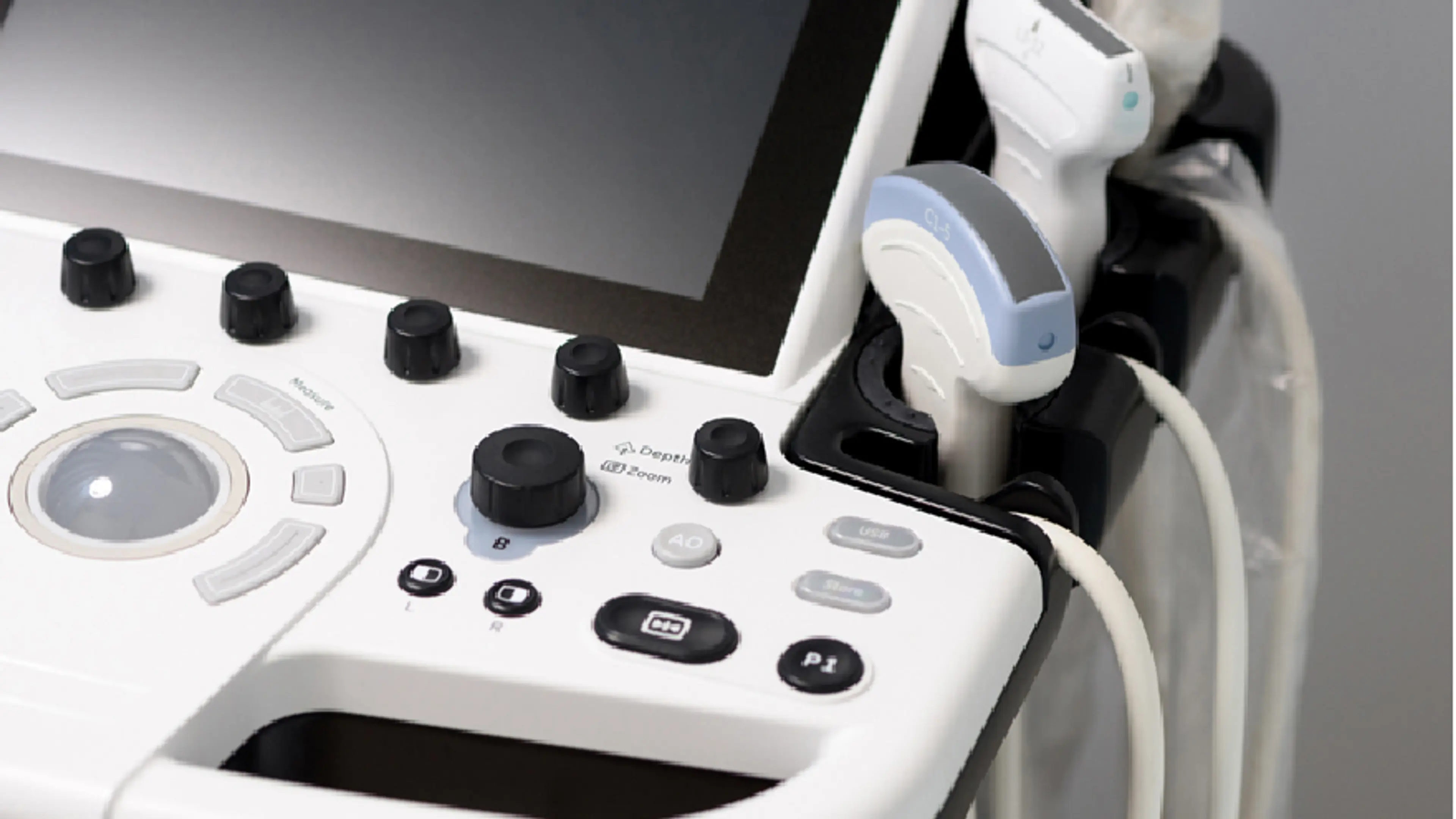
Ultrasound works by emitting high-frequency sound waves, which are beyond the range of human hearing, from a small handheld device called a transducer. These waves travel through the body and bounce off organs and tissues, creating echoes. The transducer picks up these echoes and sends the information to a computer that generates detailed visual images of the internal structures. These images can then be analyzed by healthcare providers for diagnostic purposes.
One of the most important features of ultrasound is that it is non-invasive and radiation-free, unlike other imaging methods such as X-rays and CT scans. This makes ultrasound particularly advantageous for vulnerable populations like pregnant women, children, and those needing frequent imaging. Moreover, the technology is relatively inexpensive and widely available, making it an accessible option for many medical facilities.
In recent years, ultrasound technology has expanded beyond traditional uses.
3D ultrasound provides highly detailed, three-dimensional images that allow for better visualization of organs and tissues. In
prenatal care, 3D ultrasound allows doctors and parents to see the fetus in more detail than ever before, offering insight into growth and development.
AI integration into
ultrasound technology has also improved diagnostic accuracy, allowing computers to assist in identifying potential abnormalities with greater precision than ever before.
Ultrasound in Early Disease Detection: Key to Lifesaving Interventions
ultrasound-in-early-disease-detection:-key-to-lifesaving-interventionsOne of the primary advantages of ultrasound is its ability to identify medical issues early, often before they cause symptoms. Early diagnosis significantly improves the effectiveness of treatments, as doctors can address problems before they worsen. Timely intervention can often prevent serious complications, improving patient outcomes.
Detecting Cancer Early
detecting-cancer-earlyCancer detection is a prime example of how ultrasound can save lives. Early detection of many cancers—such as breast cancer, thyroid cancer, and liver cancer—is crucial for increasing survival rates. Ultrasound is particularly effective in detecting soft tissue tumors, such as those found in the breast or thyroid, which may not be easily detected through other imaging methods like mammography or CT scans.
For instance, breast cancer is often detected with ultrasound in women who have dense breast tissue, where mammograms may miss tumors. Ultrasound can help detect abnormalities that are not visible on a mammogram, enabling early biopsy and treatment. Thyroid cancer can also be diagnosed early through ultrasound, especially for patients who may have thyroid nodules. Identifying these nodules early allows doctors to perform further testing, such as a fine-needle aspiration biopsy, to determine if the nodule is malignant.
In liver cancer, ultrasound is particularly useful in detecting tumors that may not show symptoms in the early stages. Since liver cancer often develops quietly, without noticeable symptoms until it has advanced, early detection via ultrasound can be a lifesaver. Once identified, treatment options such as surgery, ablation, or chemotherapy can be considered, which may significantly increase the chance of a favorable outcome.
Abdominal Ultrasound: Detecting Organ Issues
abdominal-ultrasound:-detecting-organ-issuesAbdominal ultrasound is one of the most commonly used ultrasound exams, providing crucial insights into the condition of internal organs, including the liver, gallbladder, kidneys, and pancreas. Several diseases affecting these organs can remain symptom-free until they have progressed significantly, making regular abdominal ultrasounds a valuable screening tool.
For example, liver disease, such as fatty liver disease, cirrhosis, and even liver cancer, can be detected early with an abdominal ultrasound. The scan can reveal subtle changes in the liver's texture or size, which may indicate a potential problem. Catching liver disease early allows doctors to intervene before the disease becomes irreversible.
Similarly, gallstones are often detected during an abdominal ultrasound, even before they cause symptoms like pain or nausea. Since gallstone disease can progress without significant symptoms until it is severe, early detection allows for minimally invasive treatments or procedures to remove the stones before they cause more severe problems.
The kidneys are also commonly examined through abdominal ultrasound, where kidney stones, cysts, or signs of chronic kidney disease (CKD) can be detected. Early diagnosis of kidney problems is essential for preventing further damage, especially in conditions like CKD, where early intervention can slow or even stop the disease's progression.
Pregnancy and Prenatal Ultrasound: Safeguarding Mother and Baby
pregnancy-and-prenatal-ultrasound:-safeguarding-mother-and-baby
One of the most well-known applications of ultrasound is prenatal care, where it is used to monitor the health of both the mother and the developing fetus. Ultrasound is invaluable during pregnancy for confirming the pregnancy, determining the due date, assessing fetal growth, and detecting potential complications early. It’s an essential tool for assessing the development of the baby and ensuring both maternal and fetal health.
Ultrasound in pregnancy can detect ectopic pregnancies, birth defects, and growth restrictions that may not be apparent through physical examination or other tests. For example, early fetal abnormalities, such as spina bifida or cleft lip, can often be detected through routine ultrasound scans. The ability to identify these issues early gives doctors and parents the opportunity to plan for interventions or specialized care at birth.
Recent innovations in 3D ultrasound technology have further enhanced the ability to monitor fetal development. These scans provide highly detailed, three-dimensional images of the fetus, allowing healthcare providers to better assess growth, anatomy, and any potential issues. Additionally, 3D ultrasounds can be used to create detailed images of the baby’s face, which can be a rewarding experience for expecting parents.
Ultrasound for Heart Health: Detecting Disease Before Symptoms Arise
ultrasound-for-heart-health:-detecting-disease-before-symptoms-ariseHeart disease remains the leading cause of death worldwide, but many heart conditions can be detected early with ultrasound, potentially saving lives. Echocardiograms, a specialized form of ultrasound, provide detailed images of the heart's structure and function. This test can identify issues such as valve defects, fluid around the heart, and early-stage coronary artery disease (CAD).
Coronary artery disease often develops without symptoms in the early stages, making it one of the most dangerous conditions. Ultrasound, particularly echocardiography, can reveal early warning signs of CAD, including plaque buildup in the arteries that restricts blood flow to the heart. By detecting CAD early, doctors can prescribe lifestyle changes, medications, and other preventive measures to reduce the risk of a heart attack.
In addition to its role in diagnosing CAD, ultrasound is also an essential tool in assessing the heart’s overall function. Regular echocardiograms can help doctors monitor patients with existing heart conditions and make timely interventions to prevent further complications.
Localized Expertise: Sangdo Woori Internal Medicine’s Use of Ultrasound
localized-expertise:-sangdo-woori-internal-medicine's-use-of-ultrasoundAt Sangdo Woori Internal Medicine Clinic, ultrasound plays a central role in the diagnostic services we provide. Led by Dr. Yoo Du-yeol, our clinic uses the latest ultrasound technology, including 3D imaging and AI-enhanced diagnostics, to offer highly detailed and accurate assessments of a wide range of health conditions.
For instance,
Dr. Yoo Du-yeol and our team have successfully identified
early-stage liver disease and
kidney stones in patients who were not yet exhibiting symptoms, helping them receive timely treatment. Additionally, we use ultrasound to monitor patients with chronic conditions, ensuring that we catch any complications early and intervene before they become severe.
We take a patient-centered approach to care, ensuring that all ultrasound results are clearly explained, and any necessary follow-up steps are taken promptly. Our commitment to using state-of-the-art ultrasound technology enables us to offer high-quality care and improve patient outcomes.
Conclusion: Why Ultrasound Should Be Part of Your Routine Health Check
conclusion:-why-ultrasound-should-be-part-of-your-routine-health-check
Ultrasound technology offers a non-invasive, radiation-free, and cost-effective way to detect a wide range of health issues early, often before symptoms appear. From heart disease and cancers to thyroid disorders, kidney disease, and organ dysfunction, ultrasound provides valuable insights into a patient's health and helps doctors identify problems when they are most treatable.
At
Sangdo Woori Internal Medicine Clinic, we are committed to leveraging the latest ultrasound advancements to provide our patients with the best possible care. Our team, led by
Dr. Yoo Du-yeol, ensures that all patients receive thorough, effective, and personalized care. Early detection through ultrasound not only helps to catch diseases in their early stages but also significantly improves the chances of successful treatment.
Get in Touch
get-in-touchAre you due for an ultrasound? Whether it’s for routine screening, prenatal care, or monitoring a specific health condition,
Sangdo Woori Internal Medicine Clinic is here to help.
Schedule an ultrasound today and take proactive steps toward safeguarding your health.