Home / Articles
What an Echocardiogram Can Reveal About Your Heart
Home / Articles
What an Echocardiogram Can Reveal About Your Heart
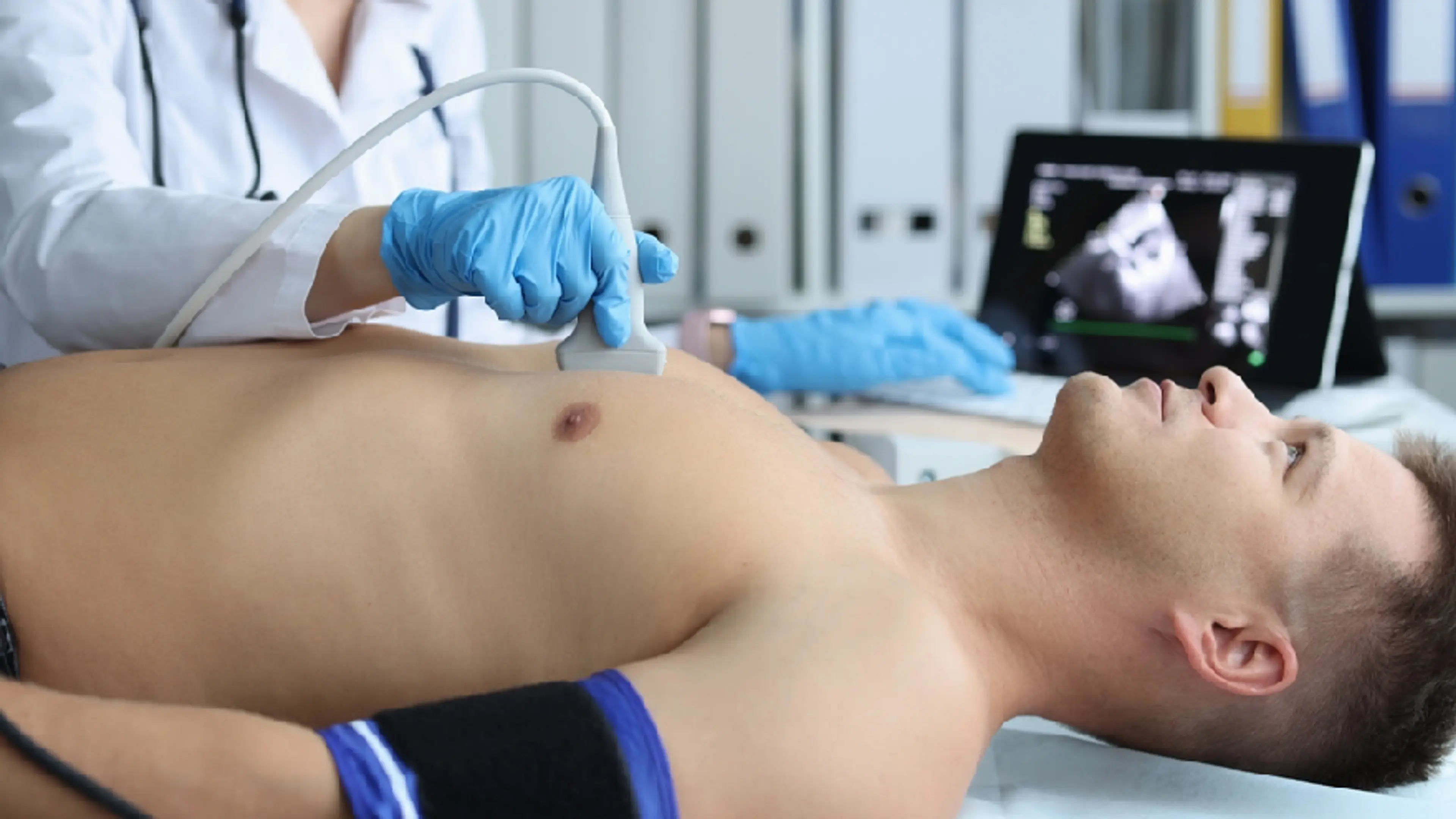
An echocardiogram uses ultrasound waves—the same technology used during pregnancy—to produce real-time images of the heart. These images show how your heart beats, how blood flows through its chambers, how the valves move, and how the muscle contracts and relaxes.
What makes an echocardiogram so powerful is that it doesn’t just take a snapshot—it shows dynamic, moving footage. Your doctor can see how your heart performs in real time, from beat to beat.
There are different types of echocardiograms (which we’ll explore shortly), but they all aim to answer one key question: Is your heart working the way it should?
One of the first things an echocardiogram reveals is whether the chambers of your heart—the left and right atria and ventricles—are normal in size and shape. If the heart muscle has thickened due to long-term high blood pressure, or if a chamber has stretched due to valve issues or heart failure, it will show up clearly.
A central metric revealed by an echocardiogram is ejection fraction, or EF. This tells us how much blood your heart pushes out with each contraction, especially from the left ventricle—the main pumping chamber.
A normal EF is between 55% and 70%
An EF below 40% may indicate heart failure or weakened heart muscle (cardiomyopathy)
An EF that is too high might suggest diastolic dysfunction or an overworked heart
Some patients walk through our doors with mild fatigue, occasional dizziness, or swelling—and find out their EF has dropped quietly over the years. In many cases, timely medication, dietary shifts, and careful monitoring can restore stability and prevent long-term damage.
Your heart has four valves that act like doors—ensuring blood moves in the right direction. If any of these valves leak (regurgitate), become too tight (stenosis), or don’t close properly, the heart has to work harder than it should.
Using Doppler ultrasound, the echocardiogram can show how blood flows through each valve and how fast. It can detect:
Dr. Yoo Du-yeol emphasizes that early detection of valvular disease often avoids surgery. In many cases, close monitoring and medical therapy are enough to preserve function for years.
Echocardiography can uncover conditions that don’t show up on an ECG or even some blood tests. For instance:
Old heart attacks: scarring or weakened areas of the heart wall
Congenital defects: small holes between chambers (ASD or VSD)
Blood clots or masses: especially in patients with atrial fibrillation
Fluid around the heart (pericardial effusion): which can impair the heart’s ability to fill and pump
These findings can explain vague symptoms like fatigue, chest pressure, or dizziness that have no obvious cause in routine tests.
Pumping isn’t the only thing that matters. The heart also needs to relax properly to refill between beats. Echocardiography can assess diastolic function—how well the heart relaxes and receives blood.
In some cases, a patient’s EF might be normal, but they still experience symptoms of heart failure. This is called heart failure with preserved ejection fraction (HFpEF), and it’s increasingly common in older adults, especially those with high blood pressure, diabetes, or thyroid issues.
Through detailed echo parameters—like E/A ratio, tissue Doppler velocity, and left atrial pressure estimation—doctors can detect this earlier and manage it effectively.
The standard, non-invasive echo
Done with a probe placed on your chest
Ideal for routine evaluation and general screening
Combines ultrasound imaging with physical or pharmacologic stress
Shows how the heart performs under exertion
Especially useful for detecting hidden coronary artery disease
Uses a small probe inserted into the esophagus
Offers clearer images of hard-to-see structures like the aorta and valves
Often used when the chest wall or lungs block clear views
Provides advanced visualization of heart valves and chamber movement
Helps in surgical planning or complex diagnosis
Dr. Yoo and our cardiology network will determine which test is most appropriate—based on your symptoms, history, and risk profile.
Echocardiography has become the cornerstone of heart diagnostics for good reason:
It’s also repeatable. For chronic disease patients at our clinic, we might repeat echos every 6–12 months to track disease progression, therapy response, or valve health.
In Korea’s aging population—especially among those in their 50s and 60s who are actively managing hypertension, thyroid disease, or metabolic syndrome—echo screenings are a smart, proactive choice.
We’ve had patients who came in with no symptoms at all, just a family history of heart disease. Their echocardiogram revealed mild valve leakage, or a stiffening ventricle, or an enlarged left atrium. Were they in danger? Not immediately. But now they have a roadmap, and we have a plan.
In another case, a patient recovering from COVID-19 experienced unexpected fatigue. A post-viral echo revealed mild myocarditis—a condition that would have gone unnoticed without imaging. Early detection meant targeted rest and follow-up, preventing long-term scarring.
That’s the power of knowing.
What We Check | What It Can Show |
|---|---|
Chamber size and wall thickness | Enlarged heart, hypertrophy |
Ejection fraction (EF) | Heart failure, pump efficiency |
Valve motion and blood flow | Regurgitation, stenosis, prolapse |
Diastolic function | Relaxation issues, HFpEF |
Structural issues | Scars, clots, tumors, congenital defects |
Fluid around the heart | Pericardial effusion, inflammation |
Response to stress | Coronary artery disease, silent ischemia |
Advanced views (TEE/3D) | Pre-surgical detail, complex anatomy |
Your heart speaks—not with words, but with motion, rhythm, and flow. The echocardiogram is how we listen.